
Fixed specimen confocal image showing labeled cholesterol vacuoles
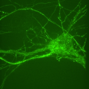
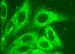
Primary neuron cultures from rat. Intradendritic transport of GFP labeled particles from time lapse sequence. Image acquired by Solamere Technology confocal microscope and Stanford Photonics XR/MEGA-10EX™ camera. 100X

Live XR/MEGA-10 ™ confocal image of macrophages showing capping of HIV protein labeled with FITCI by Dr. Bruce Freedman
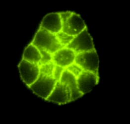
Three-dimensional culture of human mammary epithelial
cells showing FITC-labelled E-cadherin expression. Three-dimensional cell
culture systems are being developed as better models for breast cancer
initiation and progression at Lawrence Berkeley National Laboratory by
Dr. Mina Bissell. Image acquired by Solamere
Technology spinning disk confocal microscopy onto a Stanford Photonics
XR/MEGA-10EX™
camera, courtesy of Damir Sudar, Staff Scientist at LBNL.
Life Sciences Applications
Historical Perspective
Stanford Photonics began building Intensified CCD (ICCD) cameras for life science imaging in 1989. The fiber optic bonding technology essential to optimizing performance of ICCD’s had been developed and improved in prior years via ongoing projects and OEM products tied to military night vision and surveillance. The first units built for fluorescence microscopy leveraged this capability and also relied on strategic alliances with several camera manufacturers as sources of state of the art CCD’s and board level drive electronics. A number units were custom designed for a small group of researchers at the University of Maryland who had purchased a batch of high performance image intensifier tubes based on a recommendation by Ken Spring at the NIH. He had shown that a device developed by Philips Electronics specifically for military night vision could be modified and used to produce outstanding low light fluorescence images from live cells with image quality far exceeding the prior art. The image intensifier was the KS 1381. As far as we know, these first cameras are still in use today.
In the early 90’s, attempts to commercialize this product on a larger scale were thwarted by the inability of the supplier to make new KS-1381’s that looked as good as the genesis lot. Philips had “lost the process.” Other GEN II image intensifiers were evaluated and rejected. Finally, involvement in a Naval research program led to the testing and qualification of a variant GEN III with a new photocathode material specially developed and paid for by the Navy for imaging in the blue to green spectral band. As a result, in 1993 Stanford Photonics was able to introduce Gallium Arsenide Phosphide (GaAsP) technology to the life sciences. The combination of high quantum efficiency and low dark counts made this detector ideal for video rate, ratiometric Calcium imaging. Unfortunately, within a year, the company producing GaAsP tubes was acquired and the production line was shut down and dismantled. As the “last buy” lot of tubes was being shipped to customers, the search for a replacement commenced, and, again, we found another new product coming out of the military market. Stanford Photonics received prototype specifications for the first GEN III“Ultra Blue” intensifier tubes (historically referred to as “GEN IV”) at the end of 1993 and announced it’s general availability at Neuroscience, November 1995. This perseverance in finding and uncovering the newest and the best is a major component of Stanford Photonics corporate culture that continues to drive our success today.
In Summary...
Since the early ‘90’s, Stanford Photonics has consistently been the first company to test and market advancements in GEN III technology in the microscopy marketplace, including unfilmed GEN III and partially filmed “Pinnacles” with extended blue response and, most recently, the resurrection of GaAsP. This has been accomplished in part as a result of our close ties with the companies developing and the customers demanding the best in night vision. We believe that this unique synergy will continue to benefit and drive low light, live cell imaging to new horizons of discovery.
To Learn more...
Specification sheets are available for the following products:
XR/MEGA-10 ™ and XR/MEGA-10LC ™
- GEN III, megapixel
- Most cost effective
- Unfilmed technologies
- Highest performance
- Single Photon detection
- Single Molecule and Luminescence Imaging
For more outstanding images and information
about specific applications, users and installations, and ancillary products,
please visit Solamere
Technology Group .
Stanford Photonics, Inc. · 1032 Elwell Court · Suite 104 · Palo Alto · CA 94303 · Phone: 650.969.5991 · Fax: 650.969.5993 · E-mail: info@stanfordphotonics.com